Summary
- Researchers have created the first comprehensive spatial atlas of the developing human thymus, the organ primarily responsible for the production of immune cells
- This study shows that the foundation for lifelong immunity is established earlier than previously thought
- This could also help inform new T cell engineering therapies for cancer treatments, autoimmune conditions, and transplants
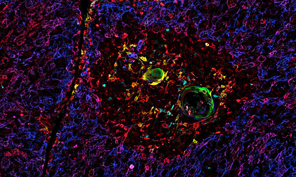
By creating the first spatial atlas of the developing human thymus, the organ that trains immune cells to protect against infections and cancer, scientists have discovered that the foundation for lifelong immunity is established earlier than previously thought.
The study, published in the journal Nature, is part of the international Human Cell Atlas (HCA) initiative to map every cell type in the human body. Insights gained from studying how thymus samples change before and after birth could help researchers generate an artificial thymus, the first step in being able to engineer therapeutic immune cells for older adults or people with compromised immune systems.
As part of this study, former EMBL’s European Bioinformatics Institute (EMBL-EBI) and Wellcome Sanger Institute Postdoctoral Fellow, Nadav Yayon, helped to develop a new standardised high resolution spatial mapping method to study various stages of thymus development
“This thymus map is the first full model of a human organ at single cell resolution and transcriptomic breadth,” said Sarah Teichmann, co-founder of the Human Cell Atlas, formerly at the Wellcome Sanger Institute, and Professor at the Cambridge Stem Cell Institute, University of Cambridge. “It represents a crucial piece of the puzzle in our effort to understand human biology cell by cell in the Human Cell Atlas. By understanding how the thymus educates immune cells from their earliest stages, we are opening up insights into immune deficiencies and autoimmune conditions.”
This paper is one of a collection of more than 40 HCA publications in Nature Portfolio journals that represent a milestone leap in our understanding of the human body.
Tracking T cell development
Despite its importance, little is known about the early development of the thymus, as it functions primarily during infancy and then gradually degenerates over the lifespan. Studying its early stages could allow us to understand why immunity wanes with age, leaving older adults vulnerable to infection and less responsive to vaccines.
In this study, researchers tracked thymus and T cell development – a type of white blood cell and part of the body’s immune response – in samples ranging from eleven weeks post-conception to three years old using single-cell sequencing and advanced spatial mapping techniques.
They discovered that the organ’s basic structure and function is established as early as twelve weeks post-conception, suggesting that early pregnancy factors may have a more profound impact on lifelong immune function than previously recognised.
Insights into the human immune system
The team uncovered key differences in the development of various T cell types; some that help orchestrate immune responses by directing other immune cells and others that directly attack infected or cancerous cells. This understanding could inform new T cell engineering therapies that selectively boost immunity for cancer treatments or suppress it for autoimmune conditions and transplants.
The researchers also discovered locations of progenitor cells that give rise to important supporting cells in the thymus which also mimic the body’s own environment so that T cells would not react to itself. This could help researchers in the future to create an artificial thymus for regenerative immune therapies for older adults or people with compromised immune systems.
OrganAxis for high resolution spatial mapping
The study also demonstrates a new standardised high resolution spatial mapping method called OrganAxis to compare the composition and structural organisation of thymus samples across various stages of development at a much higher resolution than ever before. This approach could be applied to other organs that change significantly over time or vary widely across individuals, such as the liver or kidneys.
“The thymus is uniquely crucial in setting up lifelong immunity, but until now, comparing its different developmental stages was almost impossible, as they appeared like completely different organs,” said Nadav Yayon, formerly at the Wellcome Sanger Institute and EMBL-EBI, and now Senior Computational Scientist at the Cambridge Stem Cell Institute, University of Cambridge. “OrganAxis lets us integrate different spatial datasets to uncover hidden properties that go unnoticed when viewed individually. Using key structures as reference points, much like a hiker uses landmarks to navigate, we now see how structures are formed early on, enabling us to track T cell training over time.”
This work was a collaboration between scientists from the Wellcome Sanger Institute, EMBL-EBI, Ghent University, the National Institutes of Health’s National Institute of Allergy and Infectious Diseases and others.
The full press release was originally published on the Wellcome Sanger Institute website.
Edit